Home » Without Label » Upper Thigh Anatomy - 1 : Along the upper portion of the thigh, just lateral to the gracilis, the adductor longus muscle is ranked as the most anterior of this group of thigh muscles.
Upper Thigh Anatomy - 1 : Along the upper portion of the thigh, just lateral to the gracilis, the adductor longus muscle is ranked as the most anterior of this group of thigh muscles.
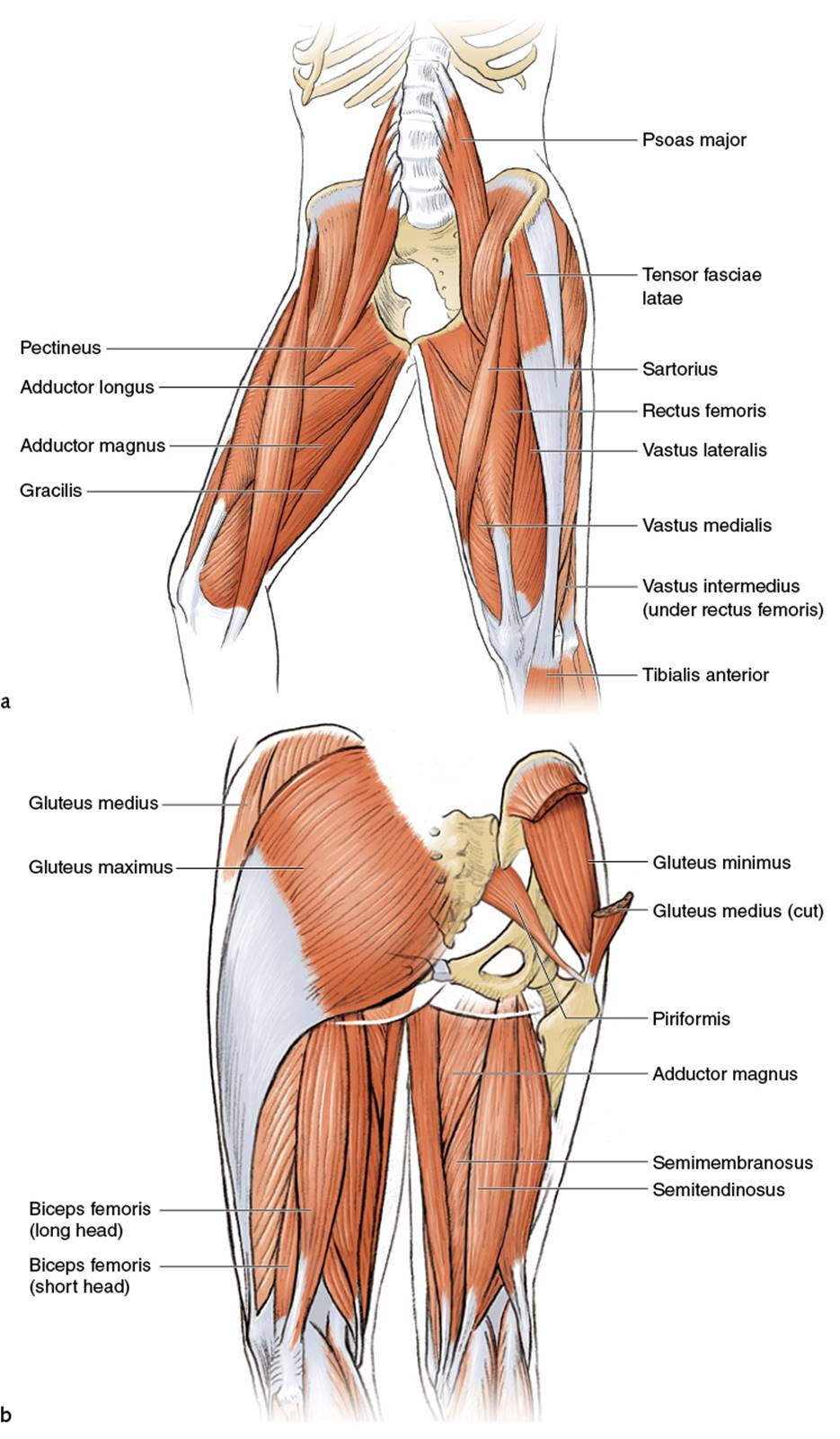
Upper Thigh Anatomy - 1 : Along the upper portion of the thigh, just lateral to the gracilis, the adductor longus muscle is ranked as the most anterior of this group of thigh muscles.. The hamstring muscles, also known as the rear thighs, make up the backside of the upper leg anatomy. Related posts of muscle anatomy of upper thigh anatomy muscle attachments. Rectus femoris, vastus medialis, vastus lateralis and vastus intermedius. The upper leg, in particular, is comprised of bones and muscles that are susceptible to injury, particularly when excess strain is placed upon them. Like the forearm, the upper leg, or thigh, has a dense arrangement of many muscles.
Upper part of the ischial tuberosity insertion: Along the upper portion of the thigh, just lateral to the gracilis, the adductor longus muscle is ranked as the most anterior of this group of thigh muscles. Anterior superior iliac spine insertion: The thigh muscles don't just move your legs. Related posts of muscle anatomy of upper thigh anatomy muscle attachments.
Anterior Surface Of The Left Thigh The Observational Area Is Download Scientific Diagram from www.researchgate.net Long bones, short bones, flat bones, and irregular bones.) long bones are longer than they are wide, with spongy bones at both ends and a cavity filled with bone. Legs give us the freedom to run, walk, jump, climb, and negotiate the world around us. It's the area that runs from the hip to the knee in each leg. Anatomy muscle attachments 12 photos of the anatomy muscle attachments anatomy muscle attachments, anatomy muscle attachments quiz, human anatomy muscle attachments, knee anatomy muscle attachments, shoulder anatomy muscle attachments, human muscles, anatomy muscle attachments, anatomy muscle attachments quiz, human. The thigh muscles don't just move your legs. Superficial fascia.—the superficial fascia forms a continuous layer over the whole of the thigh; A deep, shooting pain in the upper leg can also be caused by deep vein thrombosis, spinal stenosis, or a thigh bone infection. Spicermanyt at checkout for 40% off this tutorial!
Home remedies can often alleviate the pain, but medical treatment may also be.
There are five muscles in the anterior thigh compartment: Ebraheim's educational animated video describes muscle anatomy of the thigh. Abductors are located on the upper portion of the outside of your thighs and hips, anchoring above on the pelvis, and below at various points on your outside thigh. Biceps femoris (long head) biceps femoris (short head) semitendinosus. Along the upper portion of the thigh, just lateral to the gracilis, the adductor longus muscle is ranked as the most anterior of this group of thigh muscles upper thigh anatomy. The deep fascia is denser than its superficial counterpart and forms. There are two main types of fascia: Legs give us the freedom to run, walk, jump, climb, and negotiate the world around us. The femur is found in the thigh. Pretty self explanatory from the title i think. Home remedies can often alleviate the pain, but medical treatment may also be. The adductor brevis, adductor longus and adductor magnus make up the the starting position is lying on the right side where the upper body is supported by the right arm. Upper part of the ischial tuberosity insertion:
Any injury or disease of the hip will. Anterior muscles extend your legs and flex your thighs. 5.0 based on 12 ratings, 7 reviews. Fasciae of the hip and thigh. Rectus femoris, vastus medialis, vastus lateralis and vastus intermedius.
Leg Picture Image On Medicinenet Com from images.medicinenet.com The superficial fascia is attached to the dermis and aids in movement of the skin. In the upper thigh two distinct groups of superficial collectors were found. Experiencing pain in the inner thigh can have many causes, including a muscle strain, a hernia, and kidney stones. There are two main types of fascia: Upper inner thigh anatomy : 5.0 based on 12 ratings, 7 reviews. It is also referred to as a ball and socket joint and is surrounded by muscles, ligaments, and tendons. Anterior muscles extend your legs and flex your thighs.
Severe leg pain located around the thigh can be caused by trauma from a femoral break or muscle strain.
Anterior superior iliac spine insertion: The upper leg, in particular, is comprised of bones and muscles that are susceptible to injury, particularly when excess strain is placed upon them. By spicer mcleroy in tutorials. 5.0 based on 12 ratings, 7 reviews. The hamstring portion of the adductor magnus has a similar action to these muscles, but is located in the medial thigh. It is the largest bone in the body and is the only bone in the upper leg. Pelvic & upper thigh anatomy. Upper inner thigh anatomy : It contains many muscles and nerves but only has one bone, the femur, which is the longest and strongest bone. The femur is found in the thigh. 12 photos of the muscle anatomy of upper thigh. The muscles in the upper leg power many of our movements. Superficial fascia.—the superficial fascia forms a continuous layer over the whole of the thigh;
There are two main types of fascia: The muscles of the hip and thigh keep your hip joints strong and mighty, allowing for a wide range of hip movements. My head hurt as fuck, but whatever lmfao. The hamstring portion of the adductor magnus has a similar action to these muscles, but is located in the medial thigh. Thigh the thigh bears much of the load of the body's weight when a person is upright.
Upper Legs Running Anatomy Sports Anatomy from doctorlib.info Medial muscles adduct and rotate your thigh, and posterior flex your leg and extend your thigh. The adductor brevis, adductor longus and adductor magnus make up the the starting position is lying on the right side where the upper body is supported by the right arm. The superficial fascia is attached to the dermis and aids in movement of the skin. Rectus femoris, vastus medialis, vastus lateralis and vastus intermedius. My head hurt as fuck, but whatever lmfao. 5.0 based on 12 ratings, 7 reviews. In the upper thigh two distinct groups of superficial collectors were found. 12 photos of the muscle anatomy of upper thigh.
It is also referred to as a ball and socket joint and is surrounded by muscles, ligaments, and tendons.
The hamstring muscle attachment points. •medial thigh muscles•adductor longus muscle•adductor magnus muscle•adductor. Biceps femoris (long head) biceps femoris (short head) semitendinosus. The thigh muscles don't just move your legs. • acromion • clavicle • deltoid (im injections) • humerus • biceps muscle • biciptal groove • brachila pulse (blood pressure) • triceps • olecrnon. The superficial fascia is attached to the dermis and aids in movement of the skin. The hamstring muscles, also known as the rear thighs, make up the backside of the upper leg anatomy. It is the largest bone in the body and is the only bone in the upper leg. The upper leg, in particular, is comprised of bones and muscles that are susceptible to injury, particularly when excess strain is placed upon them. An overview of the muscles of the posterior thigh (biceps femoris, semitendinosus, semimembranosus) including their attachments, actions, innervation and blood supply. Like the adductors, the abductors are also responsible for stabilizing your knees during athletic and everyday movement. In clinical anatomy the thigh muscles are divided into three groups: The single bone in the thigh is called the femur.